

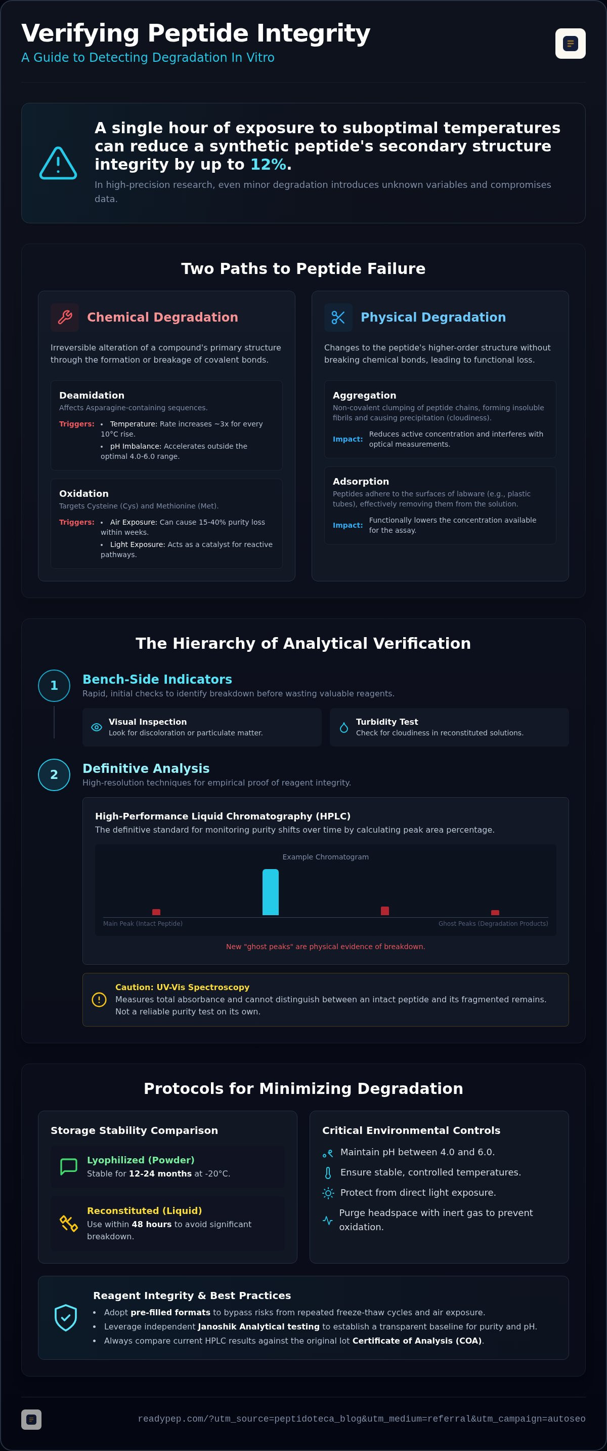
Did you know that a single hour of exposure to suboptimal temperatures can reduce the secondary structure integrity of a synthetic peptide by up to 12%? In 2026, the margin for error in metabolic research has vanished. You've likely experienced the frustration of an experiment failing due to reagent variability or shipping delays that left you questioning your compound's integrity. Detecting peptide degradation in vitro isn't just a best practice; it's a forensic necessity for data survival. When your results are on the line, "good enough" purity doesn't exist.
We understand that uncertainty is the enemy of progress. You need to know exactly what is happening in your assays. This guide provides the analytical techniques and bench-side indicators required to verify peptide stability and protect your research data. You'll learn clear protocols for identifying chemical breakdown, the critical impact of pH and temperature, and how to select reagents that minimize risk from the start. We'll cover everything from Janoshik-verified purity standards to the specific mechanics of deamidation. It's time to turn laboratory uncertainty into empirical proof.
Key Takeaways
- Differentiate between chemical pathways like oxidation and physical aggregation to accurately diagnose the root cause of inconsistent assay results.
- Implement a standardized hierarchy for detecting peptide degradation in vitro, using HPLC as the definitive standard for monitoring purity shifts over time.
- Master bench-side indicators, including visual inspection for discoloration and the "Turbidity Test," to identify breakdown before wasting high-value reagents.
- Eliminate experimental noise by adopting pre-filled research formats that bypass the risks associated with repeated freeze-thaw cycles and air exposure.
- Leverage independent Janoshik Analytical testing to establish a transparent baseline for purity and pH, ensuring every lot meets 2026 research standards.
Mechanisms of Peptide Degradation in In-Vitro Environments
Peptide degradation is the irreversible alteration of a compound's primary structure. In 2026, maintaining experimental integrity requires a forensic understanding of how these molecules fail. While a 98% purity benchmark is standard, even 1% degradation can compromise high-precision in-vitro assays by introducing unknown variables or altering binding affinities. Detecting peptide degradation in vitro starts with recognizing that a batch's purity is a snapshot in time, not a permanent state. Understanding the Mechanisms of Peptide Degradation is the first step toward securing your data against these silent failures.
Aqueous solutions are significantly more vulnerable than lyophilized powders. Water acts as both a reactant and a solvent, facilitating molecular mobility and chemical collisions. While lyophilized peptides are stable at -20°C for 12 to 24 months, reconstituted peptides in sterile water should be used within 48 hours to avoid significant breakdown. Every hour of exposure to suboptimal environments can reduce structural integrity by up to 12%, making the choice of storage format as critical as the sequence itself.
Chemical Degradation: Deamidation and Oxidation
Chemical breakdown involves the formation or breakage of covalent bonds. Deamidation is a primary concern for sequences containing asparagine. Rates of deamidation increase approximately 3-fold for every 10°C rise in temperature. Maintaining a pH between 4.0 and 6.0 is critical; levels outside this range accelerate the process. Oxidation typically targets sulfur-containing amino acids like Cysteine (Cys) and Methionine (Met). Exposure to lab air can lead to a 15% to 40% loss in purity for Methionine-containing peptides within weeks if the headspace isn't purged with inert gas. Light exposure further destabilizes peptide bonds, acting as a catalyst for these reactive pathways. Precision research compounds like GHK-Cu 100mg require strict environmental controls to prevent these specific chemical shifts.
Physical Degradation: Aggregation and Adsorption
Physical degradation involves changes to the peptide's higher-order structure without breaking covalent bonds. Surface adsorption is a "functional" form of degradation where peptides stick to the walls of plastic labware. This effectively lowers the concentration of the active compound in the assay. Aggregation is the non-covalent association of peptide chains leading to precipitation. This process often results in the formation of insoluble fibrils that alter the solution's properties. These aggregates don't just reduce concentration; they can actively interfere with optical measurements and biological responses. When detecting peptide degradation in vitro, researchers must account for these hidden losses that don't always show up on a standard visual check. Verifying the initial state via independent Janoshik Analytical reports ensures you aren't starting your research with a compromised batch.
Analytical Techniques for Quantifying Peptide Stability
In 2026, the hierarchy of analytical verification is non-negotiable. Detecting peptide degradation in vitro requires a tiered approach that moves from rapid screening to high-resolution mapping. While UV-Vis spectroscopy offers a quick tool for concentration verification, it cannot distinguish between an intact peptide and its fragmented remains. It measures total absorbance, which may remain constant even as the molecule breaks down. For researchers managing high-stakes assays, the real work begins with chromatography and mass spectrometry. These tools provide the empirical proof needed to validate reagent integrity.
High-Performance Liquid Chromatography (HPLC)
HPLC remains the definitive method for detecting purity shifts over time. By calculating the percentage purity through peak area integration, you can monitor the gradual decline of your compound. This involves dividing the area of the main peak by the total area of all detected peaks. If your original baseline shows 99% purity but your current run reveals new "ghost peaks" at different retention times, you're seeing the physical evidence of breakdown products. Comparing these current results against the original lot Certificate of Analysis (COA) is the only way to verify that your reagent hasn't compromised your data. Reliability starts with a clean baseline, and using independently tested research peptides ensures your starting purity is documented by third-party labs like Janoshik Analytical.
Mass Spectrometry and Sequence Mapping
Mass Spectrometry (LC-MS) adds the layer of identity that HPLC lacks. While HPLC tells you that a peak has shifted, LC-MS tells you why. Specific mass shifts act as chemical fingerprints for different degradation pathways. For example, a mass increase of +16 Da typically confirms oxidation, while a +1 Da shift often points to deamidation. LC-MS provides the molecular weight confirmation that HPLC alone lacks, making it indispensable for identifying specific degradation fragments. Sequence mapping goes even further, pinpointing the exact amino acid site where the chain is failing. This level of forensic detail ensures that even minor chemical alterations are accounted for before they skew your results. When detecting peptide degradation in vitro, knowing the exact molecular weight of your fragments allows you to adjust your models or discard compromised samples with confidence. Precision is the only path to reproducible data.
Red Flags: Identifying Visual and Chemical Indicators of Instability
Detecting peptide degradation in vitro doesn't always require a core facility's mass spectrometer. Often, the first signs of failure appear right at the bench. While HPLC provides the final verdict, sensory indicators and simple chemical tests serve as your first line of defense. Ignoring a slight change in solution clarity or a subtle shift in scent can lead to weeks of wasted research and irreproducible data. In the high-stakes environment of 2026 research, these bench-side red flags are just as vital as your final analytical report.
Visual and Physical Changes
Visual inspection is a mandatory pre-assay protocol. A clear solution that develops even a slight haze is likely undergoing aggregation. The "Turbidity Test" is a simple but effective method: hold the vial against a dark background with a strong lateral light source. If you see "floaters" or any particulate matter, the peptide has likely reached its solubility limit or formed insoluble fibrils. Changes in viscosity are equally concerning. If a reconstituted compound like BPC-157 + TB-500 40mg appears thicker or more "syrupy" than usual, it may indicate polymer formation. Olfactory cues are specifically relevant for sulfur-containing sequences. A sharp change in the characteristic scent of these peptides often signals oxidation before it's visible to the naked eye.
Chemical Bench-Side Testing
Chemical monitoring provides the quantitative proof needed to pause an experiment. Using micro-pH probes to verify lot consistency is a standard 2026 practice. Most peptides exhibit maximum stability in a pH range of 3.0 to 6.0. If your solution drifts toward a more basic or highly acidic state, chemical instability, such as deamidation, is likely occurring. You should also assess solubility changes upon dilution in assay buffers. If a peptide precipitates immediately upon entering a higher-pH buffer, its integrity is compromised. Documenting lot numbers and comparing bench-side results against Janoshik Analytical reports helps you track stability trends across experiments. This verification ensures that detecting peptide degradation in vitro happens before your assay begins, not after you've already collected skewed data. Consistency isn't just about the sequence; it's about the environment you maintain.
Protocols for Minimizing Degradation During Laboratory Assays
Standardizing lab protocols is the only way to ensure that the process of detecting peptide degradation in vitro leads to actionable data rather than a list of experimental failures. Prevention begins with rigorous cold-chain management. From receipt to bench-side application, any break in the thermal chain introduces structural risk. In 2026, researchers must treat thermal stability as a baseline variable. If your handling is inconsistent, your analytical results will be too.
Storage Temperature and Environment
Maintaining a consistent 2–8°C environment is essential for short-term aqueous stability. Temperature fluctuations of just 5°C can destabilize the secondary structure of sensitive compounds. If you're working with open vials, oxidation is your primary threat. Purging vial headspaces with high-purity Nitrogen or Argon (99.999%) can extend solution stability by 40% to 60%. These inert gases create a physical barrier against lab air. This prevents the oxidation of Methionine and Cysteine residues that often leads to the "ghost peaks" identified in HPLC analysis.
Handling and Aliquoting Best Practices
The "Zero Freeze-Thaw" rule is a cornerstone of modern peptide handling. Research indicates that each freeze-thaw cycle can result in a 10% to 15% reduction in active peptide concentration. While manual aliquoting is a common workaround, it increases the air-to-liquid surface area. This exposure accelerates oxidative stress and increases the risk of microbial contamination. Pre-filled research formats eliminate this trade-off by providing single-use precision without the contamination risks of manual pipetting. For comprehensive steps on maintaining reagent purity, review our research peptide pen storage guide.
Microbial degradation is another silent threat in the lab. Use strict aseptic techniques to prevent enzymatic breakdown in your in-vitro assays. Buffer selection is equally critical. Aim for physiological ionic strength while maintaining a pH between 3.0 and 6.0 for maximum stability. Photosensitive compounds like GHK-Cu 100mg require amber vials or light-shielding wraps to prevent photo-induced bond cleavage. By standardizing these handling protocols, you minimize the variables that lead to data drift. If you're looking for reagents designed to survive the rigors of high-precision research, explore the pre-filled research peptide pens available at ReadyPep.
Reagent Integrity: How Pre-Filled Formats Secure Research Data
Detecting peptide degradation in vitro is often a forensic exercise in explaining why an assay failed. Transitioning to pre-filled research formats shifts the focus from damage control to data security. Traditional vials require manual reconstitution. This step introduces ambient air, potential contaminants, and the risk of pipetting inaccuracies. Every time a vial is opened, the headspace is refreshed with oxygen. This accelerates the oxidative pathways that compromise sulfur-containing residues. Pre-filled pens bypass these variables entirely. They provide a closed system that maintains the compound's integrity from the first microliter to the last.
The Pre-filled Pen Advantage
The primary risk in manual handling is the exposure of lyophilized powder to laboratory humidity and oxygen during the reconstitution phase. Pre-filled research pens eliminate the need for manual reconstitution and the associated degradation risks. The mechanics of these pens allow for precision dosing that reduces reagent waste. It also limits the exposure of the bulk solution to the environment. This design ensures that the secondary structure remains intact throughout the study period. For a detailed breakdown of how these standards are maintained, review the ReadyPep lab-testing standards. Using a closed-loop delivery system is the most effective way to prevent the physical and chemical breakdown that skewed data in previous years.
Verification as a Baseline for Stability
Stability isn't a guess; it's a documented state. In 2026, independent testing by Janoshik Analytical is the gold standard for verifying reagent integrity. These lot reports establish your "Time Zero" purity and pH. This baseline is essential for any longitudinal study on degradation. Fragile sequences like MOTS-c 40mg are particularly sensitive to thermal stress and oxidative damage. ReadyPep utilizes global cold-chain shipping standards to ensure these compounds arrive with their chemical signatures preserved. Verification happens before the reagent enters your lab. This transparency removes the friction of uncertainty. You can focus on the data, knowing the chemistry is verified. Ensure your research integrity with Janoshik-verified ReadyPep pens and eliminate the variables that compromise in-vitro results.
Securing the Future of In-Vitro Precision
Precision in 2026 research demands more than just a sequence; it requires a verified environment. Detecting peptide degradation in vitro involves a multi-layered approach that moves from bench-side indicators to rigorous HPLC analysis. You've seen how deamidation and oxidation can silently erode data integrity, especially when using traditional multi-use vials. By standardizing handling protocols and eliminating the variables of manual reconstitution, you protect your assays from avoidable drift.
The shift toward pre-filled formats represents a significant leap in laboratory efficiency. These systems, manufactured with Metatide Healthcare precision, remove the friction of reagent preparation. Every lot undergoes independent Janoshik Analytical testing to ensure your baseline purity and pH are documented. Combined with global cold-chain shipping, this transparency ensures your research starts with a clean slate. It's time to stop troubleshooting reagent failure and start producing definitive results.
Secure your in-vitro research with pre-filled, Janoshik-tested peptide pens and maintain the high standards your data deserves. Your next breakthrough depends on the integrity of your reagents.
Frequently Asked Questions
How can I detect peptide degradation without an HPLC machine?
Bench-side indicators are your first line of defense. Use the turbidity test by holding the solution against a dark background with lateral light to spot particulates. Discoloration or a shift in scent for sulfur-containing compounds also signal breakdown. For more precision, micro-pH probes can detect drift outside the 3.0 to 6.0 stability range. While not as granular as HPLC, these methods are effective for detecting peptide degradation in vitro before starting an assay.
Does a change in pH always mean my peptide has degraded?
Generally, a significant pH shift indicates chemical instability like deamidation. Most synthetic peptides maintain maximum stability between pH 3.0 and 6.0. If your solution drifts toward a basic state, it often reflects a change in the primary structure. However, always verify that your buffer isn't the source of the drift. Monitoring pH is a reliable proxy for purity when you lack immediate access to chromatography equipment. Precision research requires this constant environmental verification.
Can degraded peptides produce false-positive results in cell culture?
Yes, degraded fragments can significantly skew in-vitro data. These breakdown products may retain partial binding affinity or trigger non-specific cellular responses, leading to false positives or unexpected toxicity. In 2026, sequence validation is mandatory for high-stakes assays because even 1% degradation introduces unknown variables. Ensuring your starting material matches the Janoshik Analytical baseline prevents these artifacts from ruining your experimental reproducibility and compromising your final results. Data integrity depends on reagent purity.
How long do pre-filled research peptide pens remain stable after the first use?
Pre-filled pens stored at 2 to 8°C typically maintain structural integrity for 25 to 30 days after the first use. This assumes the use of bacteriostatic water and a closed system that limits oxygen exposure. Unlike multi-use vials, the pen format reduces the air-to-liquid surface area. This limitation of oxidative stress is critical for preserving fragile sequences. Always refer to the lot-specific Janoshik report to verify the starting purity before beginning your usage window.
What are the most common signs of oxidation in synthetic peptides?
Oxidation primarily targets sulfur-containing amino acids like Cysteine and Methionine. Common signs include the appearance of "ghost peaks" on an HPLC chromatogram and a characteristic mass shift of +16 Da in LC-MS analysis. Visually, you might notice subtle discoloration or a change in the solution's scent. Peptides like BPC-157 + TB-500 40mg are specifically prone to these shifts if exposed to lab air. Purging vial headspaces with Argon helps prevent this chemical breakdown.
Is it possible to "rescue" a partially degraded peptide solution?
No, peptide degradation is an irreversible chemical or physical process. Once a peptide has undergone hydrolysis, deamidation, or oxidation, its primary structure is permanently altered. Physical aggregation into insoluble fibrils is also typically non-reversible in standard laboratory settings. Attempting to use "rescued" reagents will only introduce irreproducible variables into your data. It is always more efficient to discard compromised lots and replace them with fresh, Janoshik-verified reagents to ensure research integrity.
Why is cold-chain shipping critical for detecting degradation baselines?
Cold-chain shipping ensures the peptide's "Time Zero" state matches the purity documented on the Certificate of Analysis. Exposure to ambient temperatures during transit can trigger rapid deamidation or secondary structure collapse. Without a guaranteed cold chain, your baseline for detecting peptide degradation in vitro is compromised before the reagent reaches the bench. ReadyPep utilizes global cold-chain standards to prevent these transit-related failures, providing a stable foundation for metabolic research.
How do Janoshik Analytical reports help in verifying peptide stability?
Janoshik Analytical reports provide an independent, third-party verification of purity, mass, and pH for every lot. These reports establish the empirical baseline required for any stability study. By comparing your bench-side observations or subsequent HPLC runs against this initial data, you can quantify the rate of degradation over time. This transparency removes the uncertainty associated with manufacturer-only claims. It positions Janoshik documentation as the essential baseline for verifying the integrity of your research reagents.
Disclaimer
Educational content only. Not medical advice.


