
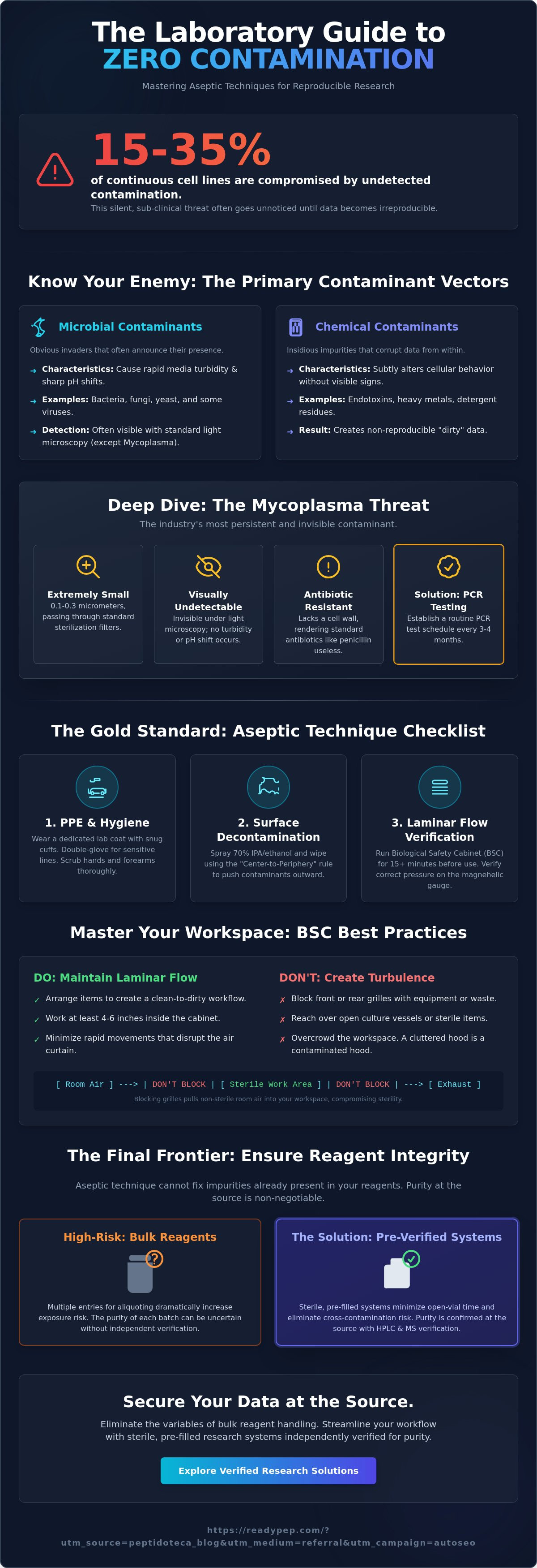
Did you know that an estimated 15% to 35% of all continuous cell lines are currently compromised by mycoplasma? It's a silent, sub-clinical infection that often goes undetected until your data becomes irreproducible. You understand the frustration of losing expensive cell lines and wasting weeks on repeated experiments due to avoidable errors. While cleaning protocols are standard, reducing contamination in cell culture requires a more rigorous focus on handling. Exposure is the enemy of precision. High standards demand a shift from reactive cleaning to proactive prevention.
This article provides the definitive laboratory checklist to eliminate microbial, chemical, and cross-contamination through advanced aseptic techniques. You will master the reagent handling protocols required to achieve zero contamination rates and highly reproducible results. We'll examine the shift toward holistic Contamination Control Strategies (CCS) and why modern labs are moving away from routine antibiotics. This guide also highlights how minimizing open-vial exposure through pre-filled research systems preserves experimental integrity. It's time to streamline your workflow and secure your data.
Key Takeaways
- Identify sub-clinical mycoplasma threats that standard light microscopy misses to protect your research against silent microbial vectors.
- Optimize Biological Safety Cabinet (BSC) setup and laminar flow efficiency to maintain a rigorous, sterile working environment.
- Discover the critical role of independent HPLC and MS verification in ensuring reagent purity and eliminating chemical contaminants at the source.
- Implement advanced strategies for reducing contamination in cell culture by transitioning away from high-risk bulk reagent handling.
- Establish a robust monitoring and documentation framework to guarantee long-term research reproducibility and streamlined laboratory workflows.
Identifying the Primary Vectors of Cell Culture Contamination
Precision in the lab starts with identifying what can go wrong. Contamination is rarely a single isolated event. It's usually a systemic failure. Effectively reducing contamination in cell culture requires a granular understanding of every vector, from the air in the Biological Safety Cabinet to the purity of your peptides. Identifying these threats before they compromise a six-month study is the difference between a successful publication and a retracted one. Exposure is the enemy of data.
Microbial vs. Chemical Contaminants
Bacteria and fungi are the most obvious invaders. They announce their presence through rapid media turbidity and sharp pH shifts. However, chemical contaminants are more insidious. Endotoxins, heavy metals, and detergent residues from improperly sterilized glassware don't kill cells immediately. Instead, they subtly alter cellular behavior. This creates "dirty" data that looks valid but lacks reproducibility. Verifying your reagent sources through rigorous lab testing is the only way to ensure chemical integrity. Maintaining a strict aseptic technique is the baseline for all in-vitro work, but it cannot fix impurities already present in your bulk reagents.
The Silent Risk of Mycoplasma
Mycoplasma is the industry's most persistent ghost. These organisms lack a cell wall, rendering standard antibiotics like penicillin entirely useless. Because they are roughly 0.1 to 0.3 micrometers in size, they stay invisible under standard light microscopy. You won't see turbidity. You won't see a pH shift. Yet, they compete for nutrients and induce chromosomal aberrations. Research from June 2026 confirms that mycoplasma continues to plague up to 35% of continuous cell lines. Relying on visual checks is a recipe for failure. Establish a routine PCR testing schedule; at minimum, every three to four months for continuous lines or whenever a new line enters the facility.
Viral and fungal vectors often exploit environmental weaknesses. Aerosols from pipetting or high humidity in incubator water pans facilitate rapid spread. Perhaps most damaging is cross-contamination between cell lines. Using the same pipette tip or reagent bottle for different cultures leads to the "hidden killer" of research. You might spend months studying a specific cell line, only to realize later that it was overtaken by a faster-growing contaminant. High standards require zero tolerance for shared reagents. Every entry into a vial is an opportunity for a breach.
The Gold Standard Aseptic Technique Checklist
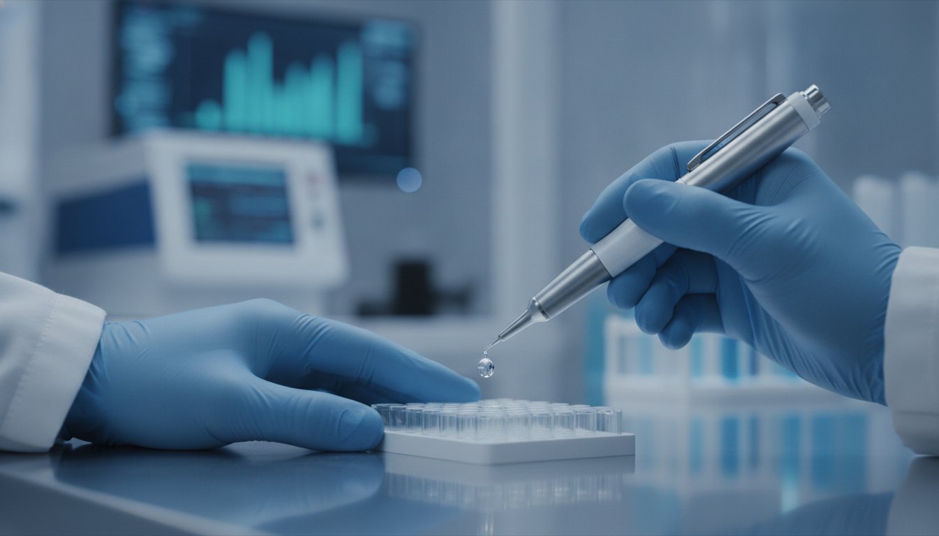
Aseptic technique is your primary physical defense. It isn't just a set of rules; it's a discipline. When reducing contamination in cell culture, your behavior inside the hood determines the validity of your results. Every movement must be calculated to preserve the sterile field. Studies on viral and bacterial contamination show that human error remains the most common entry point for pathogens. Precision begins before you even touch a pipette. Efficiency is safety. A cluttered hood is a contaminated hood.
- PPE and Hygiene: Wear a dedicated lab coat with snug cuffs. Double-gloving is recommended for high-sensitivity lines. Scrub hands and forearms with antimicrobial soap for at least 30 seconds before donning gloves.
- Surface Decontamination: Use the "Center-to-Periphery" rule. Spray 70% IPA or ethanol onto the work surface and wipe from the cleanest area (the center) toward the edges. This prevents dragging contaminants into your primary workspace.
- Laminar Flow Verification: Ensure the Biological Safety Cabinet (BSC) has been running for at least 15 minutes before use. Verify that the magnehelic gauge reflects the correct pressure for your specific unit's certification.
Optimizing the Biological Safety Cabinet
The air curtain is the only thing standing between your culture and the room's environment. Never block the front or rear grilles with notebooks, pipettes, or waste containers. This creates turbulence and pulls non-sterile air into the workspace. Arrange your equipment so you never reach over an open culture. Keep "clean" items on one side and "waste" on the other. Streamlining your research protocols with sterile, pre-filled research delivery systems reduces the number of open-vial interventions required, significantly lowering exposure risks.
In-Hood Liquid Handling Protocols
Pipetting is a high-risk activity for aerosol generation. Slow, controlled aspiration prevents the formation of micro-droplets that can contaminate the BSC's internal surfaces. Always follow the "No-Touch" rule: never let a pipette tip or bottle cap touch anything outside of its sterile packaging. If a cap is removed, place it face down on a sterile surface or hold it in a way that prevents airborne particles from settling inside. When managing multiple cell lines, process them one at a time. Clean the hood thoroughly between lines to eliminate the risk of cross-contamination. After work, run a 20-minute UV light cycle to ensure total decontamination of the interior surfaces.
Laboratory Environment and Equipment Maintenance
The laboratory environment is a living system. If the surrounding space is compromised, even the best aseptic technique inside the hood will eventually fail. Effectively reducing contamination in cell culture demands rigorous maintenance of shared equipment that often acts as a reservoir for pathogens. Cleanliness isn't a suggestion; it's a requirement for data integrity. Your environment should be as controlled as your variables.
Incubator and Environment Control
The incubator is the heart of your research. Its warm, humid interior is an ideal breeding ground for fungi and bacteria. Standard stainless steel surfaces require frequent sterilization, but implementing a copper-lined incubator strategy provides a passive, continuous antimicrobial layer. Don't rely solely on the digital readout. Perform monthly CO2 sensor calibrations using an external infrared analyzer to ensure atmospheric stability. Managing humidity is a balancing act. Use sterile, distilled water in pans and change it weekly. Stagnant water is a liability.
Shared Equipment Protocols
Communal water baths are arguably the most dangerous vectors in a modern lab. They are often overlooked hubs for Mycoplasma and fungal spores. Never submerge a bottle to the neck. If you must use a bath, treat the water with specialized antimicrobial agents and wipe every container with 70% ethanol before it enters the hood. Better yet, transition to dry-warming technologies to eliminate the liquid vector entirely. Precision in your in-vitro research protocols depends on removing these points of friction.
Centrifuges and microscopes present distinct risks. Centrifuge buckets must be sealed with aerosol-tight lids to contain potential spills or tube failures. Without these seals, a single broken tube can aerosolize contaminants across the entire room. Microscope stage plates are high-touch surfaces. They should be decontaminated after every use to prevent the transfer of microbes between plates. High-standard research requires accountability. Maintain standardized cleaning logs for all communal vortexers and centrifuges. Limit personnel traffic in the cell culture room to reduce the bioburden of airborne particles. Every breath in the room adds to the environmental load.
Reagent Integrity: Eliminating Introduction at the Source
Contamination isn't always something you pick up from the air; often, it's something you buy. Reagents are the primary vehicles for chemical and sub-clinical microbial entry. While most researchers focus on cleaning surfaces, the most significant risk to reducing contamination in cell culture often lies within the reagent bottle itself. Every time you open a bulk vial to perform an aliquot, you introduce a new opportunity for a breach. Managing reagent integrity means moving beyond faith in a label and demanding empirical verification.
Purity and Verification Standards
Purity is a verified state, not a default assumption. Independent HPLC and Mass Spectrometry (MS) testing are essential for confirming that your research peptides are free from synthesis byproducts and heavy metals. Lot-specific documentation provides the transparency required for high-standard research. Beyond microbial sterility, pH stability is a critical factor. Even minor pH variances can destabilize peptide structures or alter cellular metabolism, making your cultures more susceptible to opportunistic infections. Using reagents with documented analytical standards ensures that your baseline remains consistent across every passage.
Minimizing Handling with Pre-filled Systems
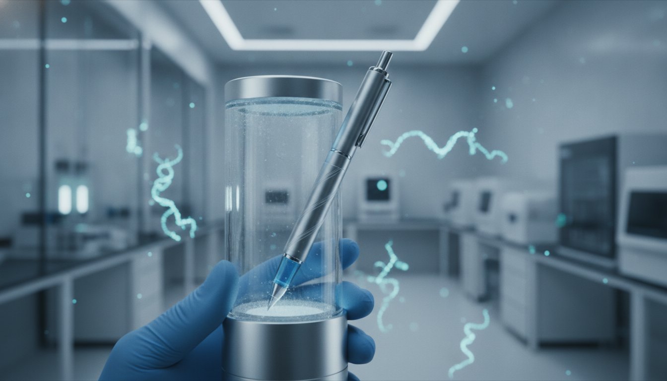
The traditional method of manual aliquoting from bulk containers is a major source of friction and error. Repeated entry into a single reagent source increases the cumulative risk of introducing airborne contaminants or cross-contaminating with other cell lines. High-throughput workflows require a more streamlined approach. Transitioning to closed-system delivery is the most effective way to maintain sterility at the source.
Practical integration of ReadyPep pre-filled pens eliminates the need for manual reagent preparation and multiple open-vial interventions. These systems provide a precise, sterile environment for your compounds until the exact moment of delivery. By reducing "open-vial" time, you effectively close the primary pathway for environmental pathogens. It's a shift from reactive cleaning to proactive containment. If you want to secure your data, you must modernize your liquid handling. Explore our full range of pre-filled research delivery systems to eliminate handling-induced contamination in your next study.
Implementation and Long-term Monitoring
Consistency is the hallmark of high-standard research. Establishing a rigorous protocol for reducing contamination in cell culture is only the first step; the second is ensuring that these standards survive the daily friction of laboratory life. Accountability must be systemic. Without a structured monitoring schedule, sub-clinical infections like mycoplasma can quietly invalidate months of data. Precision requires a shift from sporadic checks to a perpetual state of empirical verification.
Documentation and Traceability
Transparency in your sourcing is non-negotiable for publication integrity. Implementing a rigorous lot-tracking system ensures that if a contamination event occurs, you can trace the vector back to a specific batch or handling event. This level of detail is especially critical in longitudinal studies where reagent drift can alter outcomes. Understanding the nuances of peptide pen quality control allows you to maintain high-purity baselines across every phase of your research. Citing your reagent sources with lot-specific data doesn't just satisfy peer-review requirements; it protects the reproducibility of your work.
Developing a Lab Safety Culture
A sterile environment is a shared responsibility. Standardizing protocols across the entire research team eliminates the "human error" variable that accounts for the majority of breaches. Perform regular "blind" testing for mycoplasma across all active cultures. Don't wait for signs of trouble. Routine audits of aseptic technique ensure that every team member, from senior investigators to new interns, adheres to the same uncompromising standards. If a breach is detected, follow a strict disaster recovery plan. The International Society for Stem Cell Research (ISSCR) guidelines are clear: contaminated cell lines should be discarded immediately. Attempting to "cure" a contaminated culture with antibiotics often masks the issue and leads to chronic, low-level instability.
Finalizing your laboratory workflow requires a critical eye on your supply chain. High-standard research demands high-standard partners. Before starting your next passage, review your current protocols against a professional research peptide sourcing checklist. This ensures your reagents meet the analytical standards necessary for zero-contamination environments. Precision isn't accidental. It's the result of a commitment to radical transparency and technical excellence. Secure your research. Verify your source. Protect your data.
Securing the Integrity of Your In-Vitro Research
Reducing contamination in cell culture is not a one-time cleaning event. It is a continuous commitment to clinical precision. You've identified the silent threat of sub-clinical mycoplasma and mastered the center-to-periphery cleaning rule. You understand that even the most rigorous aseptic technique fails if your reagents introduce impurities at the source. Eliminating bulk-handling risks is the final step in a modern, holistic contamination control strategy. It's about removing every possible point of failure from your workflow.
Efficiency and sterility are no longer mutually exclusive. High-standard research requires tools that remove the friction of manual aliquoting and the risk of repeated open-vial exposure. Explore our independently verified research peptide pens to streamline your laboratory workflow and maintain maximum sterility. Every batch undergoes independent lot testing by Janoshik Analytical to verify purity and empirical integrity. We utilize cold-chain worldwide shipping to ensure our precision-engineered pre-filled delivery systems arrive ready for immediate, sterile integration into your study. Secure your data. Protect your results.
Frequently Asked Questions
How often should I test my cell cultures for mycoplasma?
You should test your cell cultures for mycoplasma at least once a month or immediately upon receiving a new cell line from an external source. Because mycoplasma is sub-clinical and lacks a cell wall, it evades visual detection and standard antibiotics. Research from June 2026 indicates that up to 35% of continuous cell lines are infected. Routine PCR screening is the only reliable method to confirm a sterile environment and prevent data compromise.
Can I save a cell culture once it is contaminated with bacteria?
No; you should immediately discard any cell culture showing signs of bacterial contamination. While some researchers attempt to "cure" cultures with high-dose antibiotics, this often fails and leads to chronic, low-level infections that alter gene expression. Following ISSCR guidelines, the only way to ensure research integrity is to terminate the contaminated line, decontaminate the incubator, and thaw a fresh, verified stock from your master cell bank.
What is the best disinfectant for a cell culture incubator?
Use 70% isopropyl alcohol for routine surface wiping and a non-corrosive, specialized quaternary ammonium disinfectant for deep cleaning. Avoid bleach, as it can damage stainless steel interiors and CO2 sensors. For long-term antimicrobial protection, implementing a copper-lined incubator strategy provides a passive, continuous defense against fungi and bacteria. Always use sterile, distilled water in the humidity pan and change it weekly to prevent the reservoir from becoming a vector.
How do pre-filled pens help in reducing contamination compared to vials?
Pre-filled pens help in reducing contamination in cell culture by utilizing a closed-system delivery mechanism that eliminates manual aliquoting. Traditional vials require repeated entries with a pipette, which increases the cumulative risk of airborne exposure. By using precision-engineered systems like those found at readypep.com, you bypass the need for open-vial interventions. This streamlined workflow maintains reagent sterility, securing your experimental baselines against handling-induced breaches.
Is UV light sufficient for sterilizing a biological safety cabinet?
UV light is a supplemental tool and is not sufficient as a standalone sterilization method for a biological safety cabinet. It only kills microbes on surfaces directly in the line of sight and has limited penetration depth. Shadowed areas or dust particles can protect contaminants from UV exposure. You must perform a physical wipe-down with 70% ethanol using the center-to-periphery rule before and after every session to ensure a truly aseptic workspace.
What are the signs of chemical contamination in my media?
Chemical contamination often manifests as subtle, inconsistent changes in cellular morphology or growth rates rather than obvious turbidity. You might observe altered gene expression, detached cells, or unexpected metabolic shifts caused by endotoxins, heavy metals, or detergent residues. These impurities often originate from bulk reagents. Verifying your compounds through independent HPLC and MS testing is the only way to confirm purity and eliminate these invisible variables from your in-vitro research.
Does 70% ethanol kill all laboratory contaminants?
70% ethanol is an effective disinfectant for vegetative bacteria and enveloped viruses, but it does not kill bacterial spores or non-enveloped viruses. It works by denaturing proteins and dissolving lipid membranes, which requires a specific water content to penetrate the cell wall. For comprehensive decontamination, you must supplement ethanol with sporicidal agents or autoclaving for hardware. Relying solely on ethanol leaves your lab vulnerable to persistent fungal spores and resilient microbial vectors.
How does pH affect the risk of contamination in cell culture?
Significant pH fluctuations destabilize cellular homeostasis, making cultures more susceptible to opportunistic infections. While media contains buffers like HEPES or bicarbonate, reagent impurities can cause localized pH shifts that stress the cells. This stress weakens the cell's natural resilience and can lead to faster-growing contaminants overtaking the culture. Maintaining a stable pH through high-purity reagents is a critical component of reducing contamination in cell culture and ensuring longitudinal study reproducibility.
Disclaimer
Educational content only. Not medical advice.